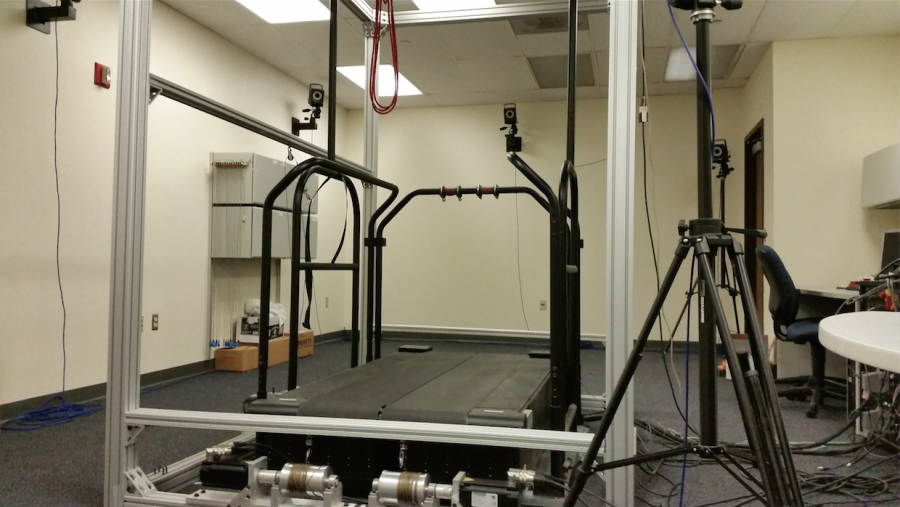
The Medical Imaging Systems Lab in the College of Engineering is a space for biomedical engineering students to focus on specific medical field research.
“Most scanners in (medical clinics) can’t use information about the energy (of the) rays that are coming in,” said Taly Schmidt, associate professor of biomedical engineering. “But it’s useful, so we’re developing ways to use that energy information to get more information about what’s going on inside (the body).”
Students experiment with scanning technology that is commonly used in medical clinics but with a twist – finding new ways to optimize it.
“We have projects that are five to 10 years out, looking at technology that’s not ready for prime time yet,” Schmidt said. He has been involved with the lab for nearly 10 years.
Working with X-ray machines, the lab develops methods to improve image quality without increasing radiation dose.
“The radiation dose carries some risk of harm, although it’s highly debated what that risk is, but there is potentially some risk for getting an X-ray or a CT scan, but the tradeoff of that is image quality,” Schmidt said.
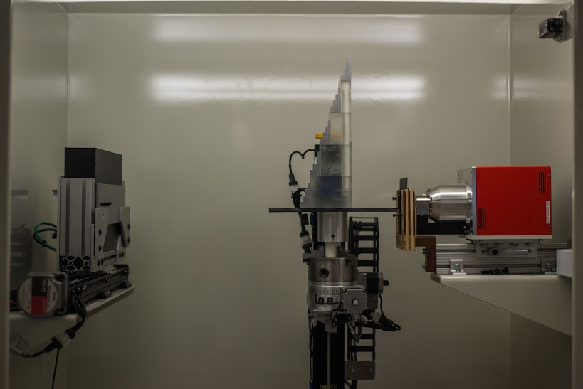
Across the street in Haggerty Hall, students who use the lab also have access to a photon-counting X-Ray detector used for research.
“At one time I could say we were one of the few (that had a detector), but the field is exploding,” Schmidt said.
Parag Khobragade, a graduate engineering student who has worked in the lab for two years, is researching ways to optimize systems to make better images.
“We’re making mathematical models to mimic the human visual system,” Khobragade said.
Kevin Zimmerman, a graduate engineering student, develops software for scanner systems to create better image quality.
“As far as a tangible product, we don’t produce any products, so we just work on algorithms and software and different methods of getting better image quality,” Zimmerman said.
Lab students don’t work together on projects unless they’re large-scale, collaborative efforts between professors and students.
One of those recent large-scale projects was Tech4POD, a pediatric research collaboration between professors from Marquette, University of Wisconsin-Milwaukee, Medical College of Wisconsin, Rehabilitation Institute of Chicago and Milwaukee School of Engineering. The professors worked in teams to address certain areas of pediatric care.
Schmidt used the lab to research fluoroscopic treatments for foot and ankle motion. She had undergraduate engineering students assist her with the work.
The definition of whether an image is “good” depends on how helpful it is to a radiologist.
“You can extract quantitative data … if the noise is low, contrast is high … but that doesn’t mean it’s a useful image to the radiologist,” Schmidt said.
The Medical Imaging Systems Lab has graduated 10 students: two Ph.D.s and eight master’s. Undergraduate students assist for shorter periods of time to help with collaborative projects like Tech4POD.
“One of the things I’m most proud of is that students that have gone through the lab and done research are now employed at GE, Siemens and Toshiba, three of the major medical imaging companies,” Schmidt said. “Marquette is well represented.”